
All of the following tutorials use the following files:
FileName: tutorial_main_planarian2.cyr
Field FileName: main_planarian2.png
The code will not be analyzed line-by-line, as most of the concepts used have been the subject of previous tutorials. Rather, we will focus on how CYCELL can model some very interesting phenomona found in flatworms.
There is an excellent summary of the notable aspects of flatworm regeneration in The Cellular and Molecular Basis for Planarian Regeneration by Peter W. Reddien. We at CYCELL have found the Field model can replicate many of these phenomona.
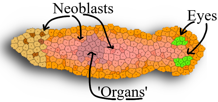
Our model flatworm is made using three types of cells: Neoblast, Body, and Eye.
We have made the outer cells a bit darker for aesthetics.
The eyes -- colored green -- are comprised of a separate type of cell from the Body and Neoblast cells that make up the rest of the worm.
The flatworm also has some organs in its middle with a purpleish pigment, as well as a tail-section that is more lightly colored. These organs and cells are Body cells that have merely been given different pigments.
Placing a few Neoblast cells next to one anther will cause a full-size flatworm to grow. A quicker way is to place a single Quickstart cell; it will quickly reproduce to make the basic body-shape and turn to the approrpriate type of cell when the growth is completed. (The trigger for the completetion of growth is when the Quickstart cells make it into the Tail region.)
This video shows basic regeneration. Various parts are removed, and they quickly grow back.
If we were to chop off the head, it would stand to reason that the worm is unable to eat (at least until it grows a new head.) Without food, regeneration must occur with existing resources. Missing tissues thus cannot be regrown to their original scale.
Our model worm simulates this phenomenon.
We will assume that the Eye cells are consistent with a mouth. That is, if the worm does not have any Eye cells, it has no mouth, and visa versa.
In the video, when we amputate the tail section, the Neoblasts very quickly run out of energy as there is no eye/mouth supplying energy. When this occurs, they will turn a darker color.
As no new cells are being made, the fragment looks dead. But, it's not... rather, the scale component of the Presumed Positions is adjusting itself, getting smaller and smaller. Eventually, a dormant Neoblast will find itself in the Field associated with the Eyes. When this occurs, it creates an eye, energy begins to flow, and the worm slowly begins growing to its full size and normal proportions.
What to look for in the video:
This video demonstrates that after parasagital amputation, the worm has no problem re-establishing its media-lateral axis. That is, there is nothing particularly vital about the axis of amputation.
We can also remove the mid-section and fuse the head and tail stubs together. You will observe how quickly new -- smaller -- organs are formed where they ought to go. The proportions of the worm are quickly modified to ensure the entire body-plan is accounted for.
Everything on this webpage as been leading up the following video.
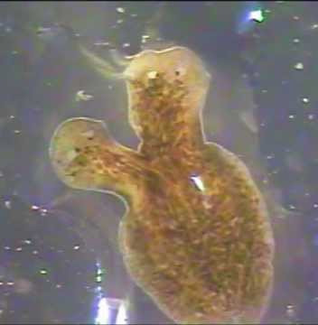
Indeed, the famous "split head" phenomona is what got us at CYCELL thinking about morphogenesis in the first place.
Upon making an incision down the head, the worm will quickly heal itself. HOWEVER, if we make an incision and then place an OBSTRUCTION the most amazing thing occurs... as in nature, the flatworm will make a duplicate head!
What to look for in the video: